You use your teeth every day for eating, speaking, and smiling, but how much do you actually know about what is inside them? From the outside, a tooth may look like a simple piece of hard material.
Under the surface, however, it is a surprisingly complex structure made of several tissues, each with a different job. Understanding the basic anatomy of your teeth can make dental care feel less confusing.
It explains why a small cavity may eventually cause severe pain, why exposed tooth roots feel sensitive, and why dentists sometimes recommend fillings, crowns, or root canal treatment.
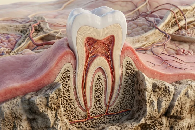
Your teeth are also different from ordinary bones. Although both contain minerals, teeth are built from enamel, dentin, cementum, and living pulp tissue. They are supported by the gums, periodontal ligaments, and jawbone.
Learning how these parts work together will help you understand your dental health and make better decisions about protecting your smile.
The Three Main Parts of a Tooth
A tooth can be divided into three main areas: the crown, neck, and root. Each area has a specific position and function within the mouth.
1. The Crown
The crown is the visible part of the tooth above the gumline. Its shape depends on the type of tooth. Front teeth have thinner cutting edges, while back teeth have broader chewing surfaces with raised points called cusps.
The crown is covered by enamel, which protects the softer tissues underneath. Although enamel is extremely strong, it can still be damaged by decay, acidic foods, teeth grinding, or physical injury.
2. The Neck
The neck, sometimes called the cervical area, is the narrow section where the crown meets the root. It is usually located around the gumline.
Plaque can easily collect in this area, especially when brushing is rushed or inconsistent. Gum recession may expose the neck and root surface, leading to sensitivity when drinking something cold or eating sweet foods.
3. The Root
The root is the part below the gumline that extends into the jawbone. Most front teeth have one root, while premolars and molars may have more than one.
The root holds the tooth securely within its socket. Inside it is a narrow root canal containing pulp tissue, nerves, and blood vessels.
Enamel: The Protective Outer Layer
Enamel covers the visible crown and acts as the tooth’s first line of defense. It is made mostly from tightly packed mineral crystals and is considered the hardest tissue in the human body.
However, being hard does not make enamel indestructible. Acids produced by plaque bacteria can remove minerals from its surface. Frequent exposure to sugary or starchy foods gives these bacteria more opportunities to produce acid.
Early mineral loss may appear as a white spot on the tooth. At this stage, fluoride and saliva may help replace some of the lost minerals. Once the enamel develops a physical hole, however, the damaged area usually requires professional treatment.
Enamel does not contain nerves, so early damage may be painless. This is one reason a cavity can develop without obvious symptoms.
Dentin: The Sensitive Layer Beneath Enamel
Dentin is located directly beneath the enamel in the crown and beneath the cementum in the root. It forms most of the tooth’s internal structure and supports the enamel above it.
Unlike enamel, dentin contains thousands of microscopic channels called dentinal tubules. These channels communicate with the living pulp in the center of the tooth.
When enamel becomes thin or gums recede, dentin may be exposed. Hot, cold, sweet, or acidic substances can then stimulate these tiny channels, producing a short, sharp sensation.
This explains why someone with gum recession may suddenly find ice water uncomfortable. Tooth sensitivity is not always caused by a cavity; worn enamel, exposed roots, aggressive brushing, and cracked teeth may also be responsible.
Because dentin is less resistant to acid than enamel, decay can spread more quickly once it reaches this layer.
Dental Pulp: The Living Center of the Tooth
At the center of each tooth is the dental pulp. This soft connective tissue contains blood vessels, nerves, immune cells, and cells involved in producing dentin.
The pulp provides nutrients to the tooth and allows it to sense pressure, temperature, and pain. It also has a defensive role, responding to irritation by producing additional dentin in some situations.
When decay moves through the enamel and dentin, bacteria may eventually reach the pulp. The tissue can then become inflamed or infected, causing persistent pain, sensitivity that lasts after a hot or cold drink, or discomfort when biting.
If the pulp is permanently damaged, a dentist may recommend root canal treatment. During this procedure, the infected tissue is removed, the inside of the tooth is cleaned, and the space is sealed.
The tooth can remain functional because it continues to receive support from the surrounding tissues.
Cementum and the Tooth Root
Cementum is a mineralized tissue covering the outside of the tooth root. It is not as hard as enamel, but it performs an essential structural role.
Tiny fibers from the periodontal ligament attach to the cementum. These fibers connect the root to the surrounding socket and help hold the tooth in position. Cementum can also contribute to limited repair around the root surface.
When gums recede, cementum may become exposed. Because it is softer and less resistant than enamel, the root surface can be more vulnerable to wear and decay.
This is why brushing harder is not necessarily better. Gentle brushing with a soft-bristled toothbrush can remove plaque without unnecessarily irritating the gums or wearing exposed root surfaces.
The Structures Supporting Your Teeth
Teeth do not simply sit inside the gums. They are supported by a group of tissues known as the periodontium, which includes the gums, periodontal ligament, cementum, and alveolar bone.
The gums form a protective seal around each tooth. Healthy gums are usually firm and fit closely around the teeth. Persistent bleeding, swelling, or tenderness may indicate inflammation and should not be ignored.
The periodontal ligament is a thin layer of connective tissue between the root and jawbone. It anchors the tooth while allowing a tiny amount of movement that helps absorb pressure during chewing.
The alveolar bone forms the sockets that hold the roots. Advanced gum disease can damage both the ligament and this supporting bone, which may eventually cause teeth to loosen.
The Four Main Types of Teeth
Most adults can have up to 32 permanent teeth, although some people have fewer because their wisdom teeth never develop, remain impacted, or have been removed. Children usually have 20 primary teeth.
1. Incisors and Canines
Incisors are the flat, sharp teeth at the front of the mouth. Their main purpose is to bite and cut food into smaller pieces.
Canines sit beside the incisors. Their pointed shape helps grip and tear food. They also help guide the upper and lower teeth when the jaw moves from side to side.
2. Premolars and Molars
Premolars are located between the canines and molars. Their raised surfaces help crush and break down food before it reaches the back of the mouth.
Molars have broad chewing surfaces designed for grinding. The third molars are commonly called wisdom teeth and are positioned at the back of each dental arch.
The different shapes are not accidental. Each type performs a specialized task, allowing the entire set of teeth to work as one efficient chewing system.
How Tooth Anatomy Guides Daily Dental Care
Knowing the structure of a tooth makes basic oral hygiene easier to understand. Brushing removes plaque from the enamel and gumline, while floss or interdental brushes clean areas that ordinary toothbrush bristles cannot reach.
Fluoride toothpaste strengthens enamel and supports the remineralization of early decay. Brushing twice daily, cleaning between the teeth, limiting frequent sugar exposure, and attending dental examinations can help prevent damage from reaching the deeper tooth layers.
Pay attention to symptoms such as persistent sensitivity, pain when biting, swollen gums, visible cracks, or dark areas on a tooth. These signs do not always reveal which layer is affected, so a professional examination may be necessary.
Understanding the basic anatomy of your teeth shows that every tooth is much more than a hard white object. Enamel protects the crown, dentin forms the main structure, pulp keeps the tooth alive, and cementum helps attach the root to its supporting tissues.
The gums, periodontal ligaments, and jawbone work together to keep everything stable. This anatomy also explains why dental problems can become more serious when left untreated. Damage may begin quietly in the enamel before spreading into sensitive dentin or living pulp tissue.
Protect these structures by brushing gently with fluoride toothpaste, cleaning between your teeth, reducing frequent sugar exposure, and visiting a dentist regularly. Start paying attention to your teeth before they hurt-not only after a problem appears.